|
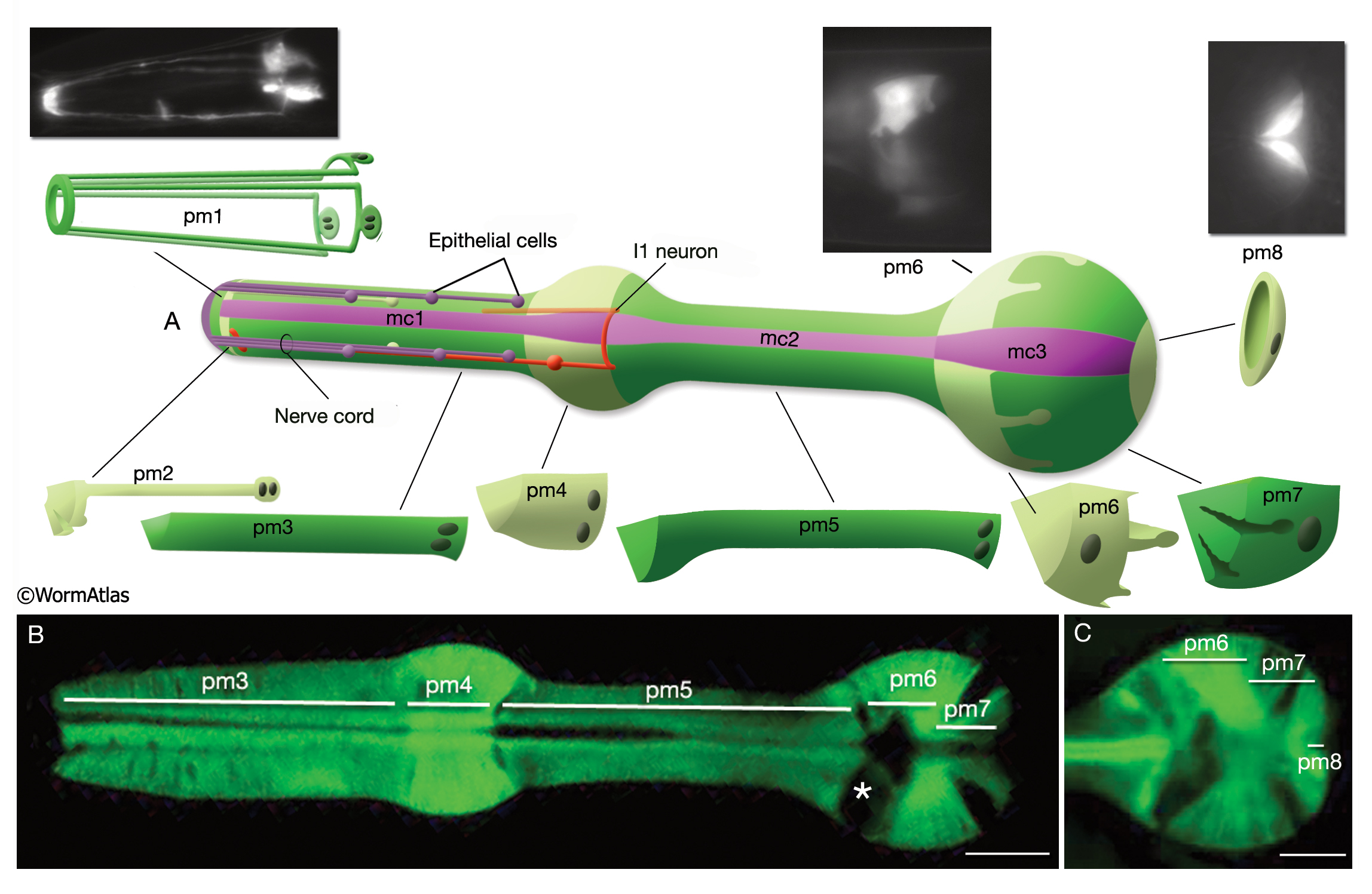
PhaFIG 6: Muscle cells of the pharynx.
A. There are eight muscle segments (pm1-pm8) in the pharynx. All but pm6, pm7 and pm8 form syncytia. The most anterior segment, pm1, is a single cell with six nuclei. Its processes extend anteriorly adjacent to the outer edges of the marginal cells and terminate in a ring structure. Each pm2 soma has two nuclei and projects into a nerve ring to extend anteriorly. pm3 cells are located in procorpus, pm4 cells in the anterior bulb, and pm5 cells in isthmus. The terminal bulb contains pm6-pm8. The last muscle segment contains a single cell, pm8, that has a single nucleus located on the left side. (Top insets) Epifluorescent images of pm1 (left), pm6 (middle), and pm8 (right) in animals expressing GFP-tagged transgenes. (Strain source: Z-W. Wang and B. Chen.) Each magnification, 400x .
B. Pharyngeal muscle cells contain radially oriented filaments. Here, myofilaments in pm3- pm7 can be seen in an epifluorescent image taken from a transgenic animal expressing the reporter gene, C32F10.8::GFP. (Asterisk) Position of g1 gland cells. (Image source: R. Newbury.)
C. Muscles of the terminal bulb. Epifluorescent image from a transgenic animal expressing the reporter gene, K02A4.1::GFP. (Image source: R. Newbury.)
Click on picture for full resolution image.
|