|
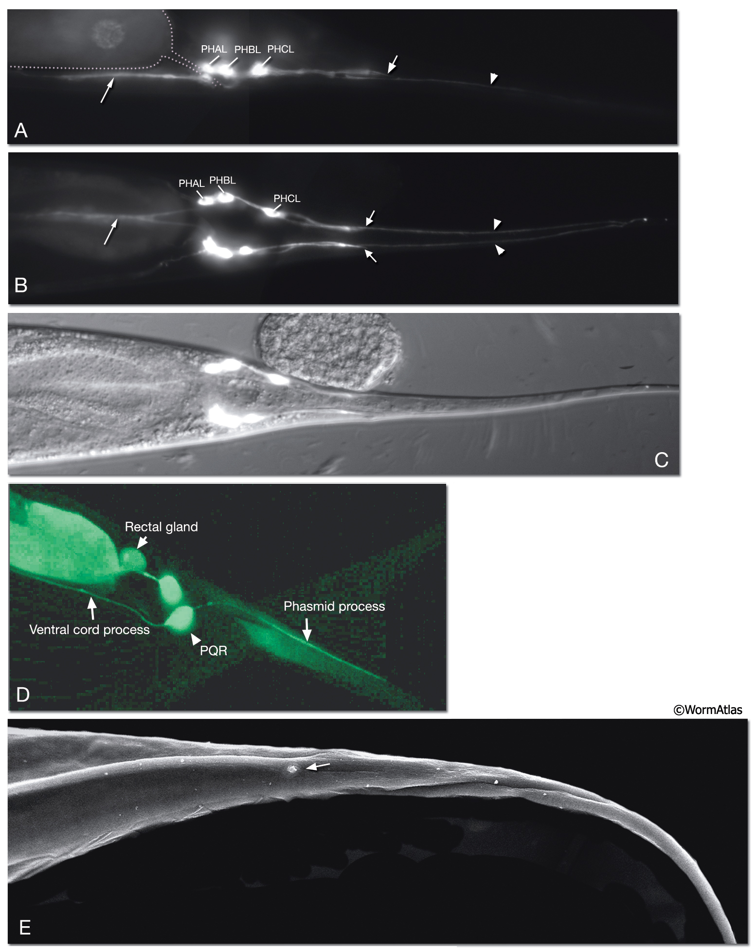
NeuroFIG 40: Sensilla of the tail tip.
The phasmids are similar in organization to amphid sensilla, but are smaller. They are located at the lateral sides of the tail and are composed of the ciliated dendrites of PHA, PHB, and, on the left side, PQR neurons as well as one sheath cell and two socket cells. Additional sensory cells extend dendrites into the extreme tail tip, but they lack cilia.
A. Epifluorescent image of a transgenic animal expressing the reporter gene pida-1::GFP, left lateral aspect. The sensillar PHA, PHB neurons, and the PHC neuron, which is not part of the phasmid, are located within the lumbar ganglia on both sides of the tail behind the rectum. (Small arrow) Phasmid openings; (arrowhead) long posterior process of the PHC neuron that extends into the tail tip. All three neurons send anteriorly directed axons (thin arrow) into the PAG neuropil via lumbar commissures. Magnification, 400x. (Strain Source: T. Zahn and J. Hutton.)
B. Epifluorescent image of a transgenic animal expressing the same reporter gene as in A, ventral view. The axons of the PHC and the phasmid neurons PHA and PHB terminate within the PAG neuropil (thin arrow) where they make synapses. The dendrites of PHA and PHB extend into the phasmid sensilla located on the lateral sides of the tail (arrows) in a symmetric fashion. These neurons modulate chemorepulsion behavior (Hilliard et al., 2002). (Arrowheads) Tail tip processes from PHC neurons, which are suggested to be proprioceptive. Magnification, 400x.
C. DIC image of the same animal as in B transposed over the epifluorescent image to indicate cell positions.
D. Epifluorescent image from a transgenic animal expressing the reporter gene ZC53.7::GFP in the postembryonic PQR neuron, left lateral view. The PQR cilium is part of the left phasmid. The PQR axon extends into the PAG via the left lumbar commissure. (Image source: R. Newbury. The Genome BC C. elegans gene expression consortium [McKay et al., 2004].)
E. SEM of an animal from the left lateral aspect showing the left phasmid sensillar opening (arrow). (Image source: D.H. Hall and C. Marks.)
Click on picture for full resolution image.
|