|
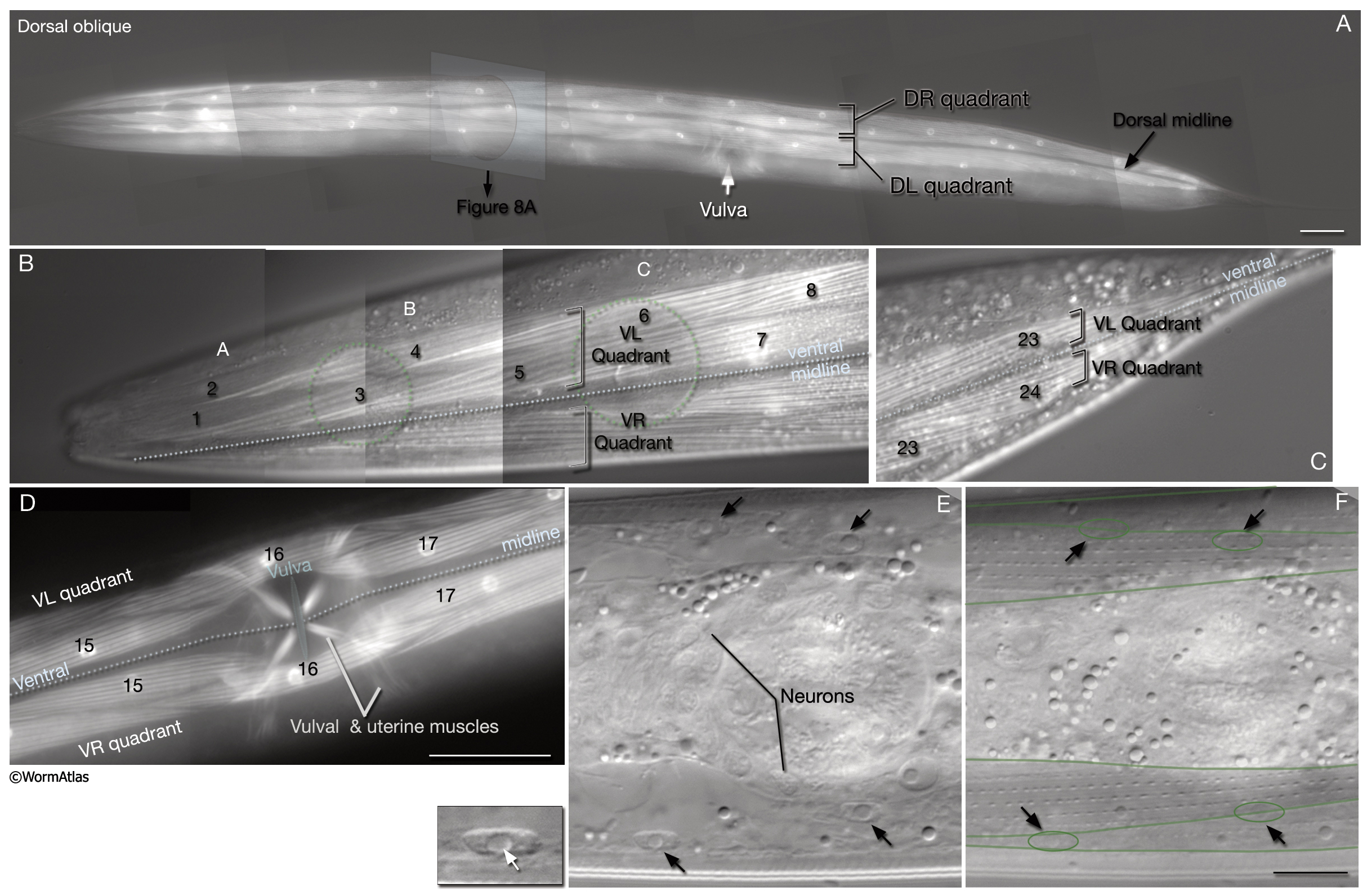
MusFIG 7: Head, neck and body wall muscles.
A-D. Epifluorescent images from transgenic animals expressing the unc27::GFP reporter gene. Muscle cells are numbered according to Hedgecock et al., 1987. (Strain source: L. Jia and S.W. Emmons.)
A. The organization of somatic muscles in the adult C. elegans hermaphrodite, dorsal oblique view. The body wall muscles are organized in four quadrants (only the dorsal quadrants are visible) with two rows of cells in each. The quadrants are placed subdorsally and subventrally. The two dorsal quadrants flank the dorsal hypodermal ridge and the dorsal cord, and the ventral quadrants flank the ventral hypodermal ridge and the ventral nerve cord. Anteriorly, the spindle-shaped cells in each quadrant are arranged almost in pairs, whereas more posteriorly the cells are organized in an alternating fashion. As a result of this arrangement, two thirds of all somatic muscle cells are located anterior to the vulva. For most cells, the muscle nucleus is centered along the anterior–posterior axis with respect to its spindle-shaped collection of sarcomeres. At the tail tip, the last muscle cell in the left dorsal quadrant continues further posteriorly in a more medial position, whereas the right dorsal quadrant terminates (not shown). Bar, 50 µm.
B. Arrangement of head and neck muscles, ventral view. The first muscle cells that are close to the midline in each quadrant are smaller than their counterparts (compare muscle cell 1 to muscle cell 2). (Green dotted lines) Positions of the anterior and posterior bulbs of the pharynx; (blue dotted line) ventral midline.
A–C correspond to the first three blocks of head muscles in quadrants. Original magnification, 600x.
C. Arrangement of the somatic tail muscles, ventral view. Original magnification, 600x.
D. Arrangement of body wall muscles near the vulva, ventral view. Bar, 50 µm.
E. Differential interference contrast (DIC) image of the posterior head in an adult animal, lateral view. Neck muscle nuclei (arrows) in the dorsal and ventral left muscle quadrants are arranged as tandem pairs. (Left inset) One of the nuclei as magnified. Smooth nucleoplasm and nucleolus (white arrow) are easily visible.
F. DIC image. Same animal as in E, but with more lateral focal plane. The boundaries of muscle cell spindles and muscle nuclei (colored with green lines) are visible. The cell bodies are arranged in tandem, similar to the nuclei. Because the nuclei lie in the more medial portions (muscle belly) of the muscle cells where the muscles are wider, the nuclei positions look somewhat skewed in this image with respect to the superficial sarcoplasmic portions. Bar, 10 µm.
Click on picture for full resolution image.
|