|
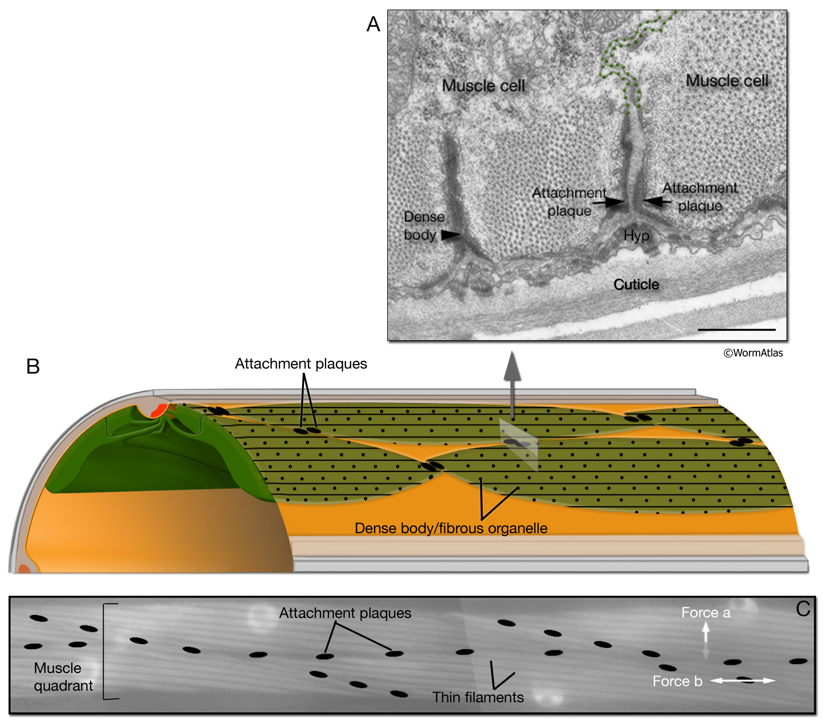
MusFIG 13: Structure of attachment plaques.
A. Attachment plaques are localized at the end of half I bands (thin filaments), where two muscle cells appose each other (green dotted lines indicate muscle cell membranes). Ultrastructurally, attachment plaques resemble DBs as electron-dense finger-like structures, and they also share some of the protein components of DBs. TEM, transverse section. Bar, 0.5 µm. (Image source: N2Y [MRC] 689-2/794L.)
B. Schematic illustration of localization of attachment plaques between the muscle cells of the dorsal left somatic muscle quadrant.
C. Placement of attachment plaques between two rows of a muscle quadrant. I bands are seen as light, longitudinal bands within each cell. Plaques are pseudocolored over an epifluorescent image taken from a transgenic animal expressing the unc-27::GFP reporter gene. Attachment plaques relay some tension longitudinally between cells (force b), but the major force generated by muscle contraction is transferred to the cuticle through basal attachments and FOs that are distributed along the entire length of the cell (force a, which acts orthogonally to this focal plane).
Click on picture for full resolution image.
|