|
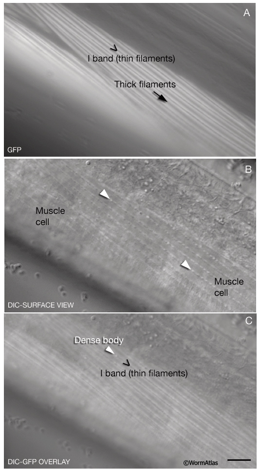
MusFIG 12: Surface views of obliquely striated somatic muscles of C. elegans.
All images are from a transgenic adult animal expressing the unc27::GFP reporter gene. Bar, 10 µm. (Strain source: L. Jia and S.W. Emmons.)
A. Dorsal view of two staggered spindle-shaped cells (one is partly out of the plane of focus) from a single quadrant. Epifluorescent image. I bands are labeled with the troponin-GFP marker, and each I band appears as a pair of longitudinal white lines. A bands (thick filaments) are not labeled and their territories appear dark. The border between the two muscle cells is demarcated by a disruption in the linear pattern.
B. The same plane of focus as in A (DIC image). DBs (arrowheads) are visible as rows of small bumps organized in the same orientation as the myofilaments. M lines, which occupy the middle of A bands, are smaller and not clearly visible.
C. Overlay of the images in A and B. DBs occupy the middle of the I bands.
Click on picture for full resolution image.
|