|
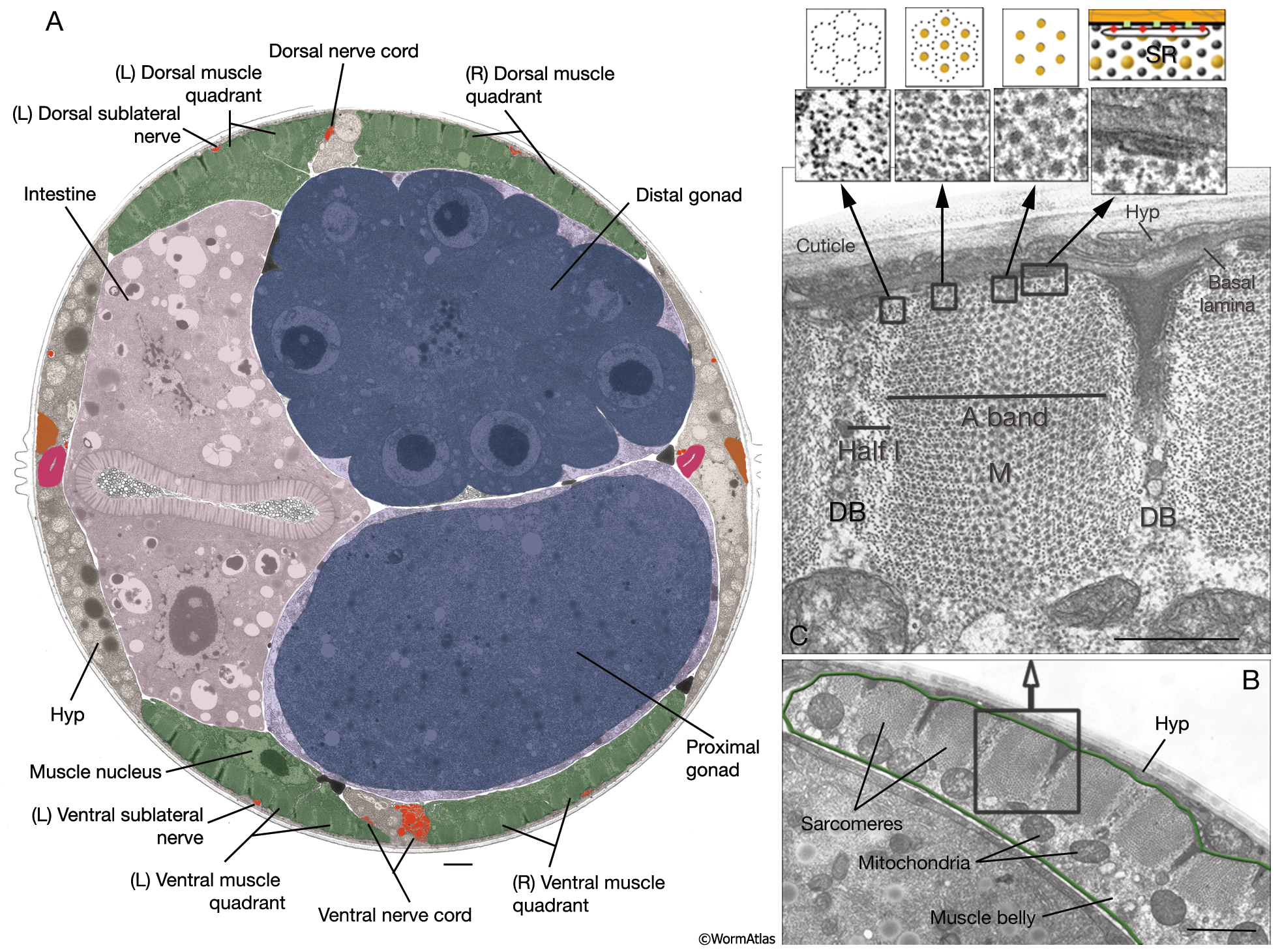
MusFIG 10: Structure of the C. elegans body wall muscle cell.
A. Low-power TEM showing the relationship of the muscle quadrants to the hypodermis and internal organs, transverse section. Bar, 1 µm. (Image source: [Hall] N501-N354.)
B. Sarcomeres in a higher-magnification TEM, transverse section. Each adult hermaphrodite muscle cell may grow to be as wide as 10 sarcomeres, which are the repeating contractile units underneath the muscle cell plasma membrane facing the hypodermis (Hyp). Mitochondria cluster at the boundary of the myofilament lattice and the muscle belly. The muscle belly also contains the nucleus and other organelles. Bar, 1 µm. (Image source: [Hall] N501C.)
C. Cross section of a single sarcomere. Same image as in B, magnified. Parts of two DBs are seen at the ends of half I (thin filament) bands on each side. A thin M line is seen to occupy the middle of the A (thick filament) band. The membranous sacs of sarcoplasmic reticulum (SR) align around the dense body and are also present under the thick and thin filament bands along the muscle membrane (top right inset). The arrangements of thick and thin filaments are shown in the top insets on the left, as both magnified TEM images and schematic drawings. Muscle cell is separated from the hypodermis and cuticle by the basal lamina (orange layer). Bar, 0.5 µm.
Click on picture for full resolution image.
|