|
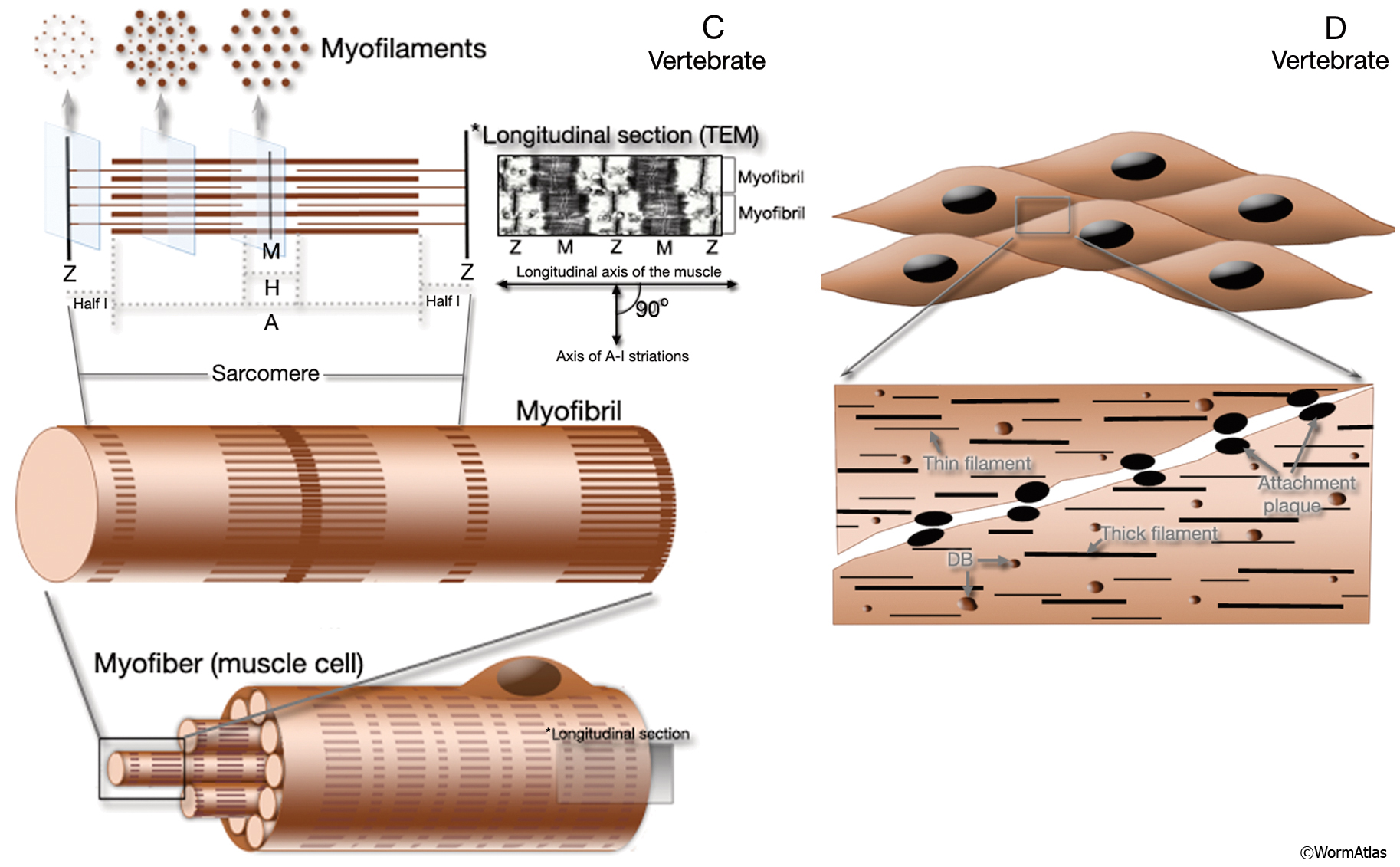
MusFIG 1C&D: The contractile apparatus in vertebrates.
C. Diagram illustrating vertebrate striated muscle. Vertebrate somatic muscle is comprised of numerous multinucleated myofibers, each of which contains many contractile myofibrils with repeating sarcomeres and develops by fusion of embryonic myoblasts. A single contractile unit between two Z discs in each myofibril is a sarcomere. In each sarcomere, myosin-containing thick filaments (thick brown bands) are interdigitated with actin-containing thin filaments (thin brown bands) on either side. Thin filaments are attached end to end at the Z lines, in the middle of the I-bands, and thick filaments are attached end to end at the M-lines, in the middle of the A-bands. Muscle contraction involves myosin sliding past actin to shorten the sarcomere.
D. Diagram illustrating vertebrate smooth muscle. Vertebrate smooth muscle consists of unfused, spindle-shaped individual cells, each with a single nucleus. There is no apparent organization of the actin and myosin filaments into discrete contractile units.
See also MusFIG 1A&B.
Click on picture for full resolution image.
|