|
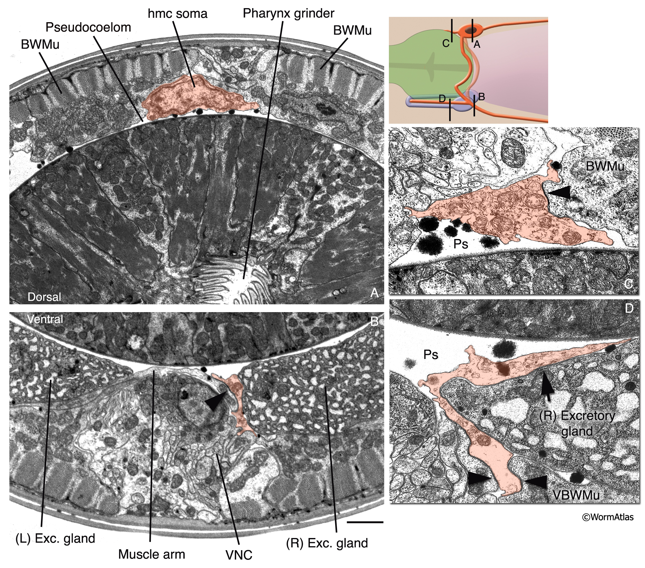
HmcFIG 3: Fine structure of the head mesodermal cell.
The hmc and hmc processes are pseudocolored as transparent orange on transverse TEM sections. (Arrowheads) Gap junctions. Bar, 1 μm.
A. Cross section of the hmc soma, which is located within the pseudocoelom ventral to the dorsal hypodermal ridge, at the level of the pharyngeal grinder. (Image source: N2U [MRC] A230-20.)
B. Ventral posterior process of hmc is located in close association to the right-side excretory gland soma at this level and makes gap junctions to ventral muscle arms (arrowhead). (VNC) Ventral nerve cord. (Image source: N2U [MRC] 240-18.)
C. The short, dorsal anterior process of the hmc makes a gap junction to a dorsal body wall muscle. (Ps) Pseudocoelom. (Image source: N2U [MRC] 245-3.)
D. The ventral anterior process of hmc runs in close association with the right-side excretory gland process. (Arrow) Possible gap junction between the hmc and excretory gland. (Image source: N2U [MRC] 235-12.)
Click on picture for full resolution image.
|