|
HypFIG 10: Anterior hypodermis.
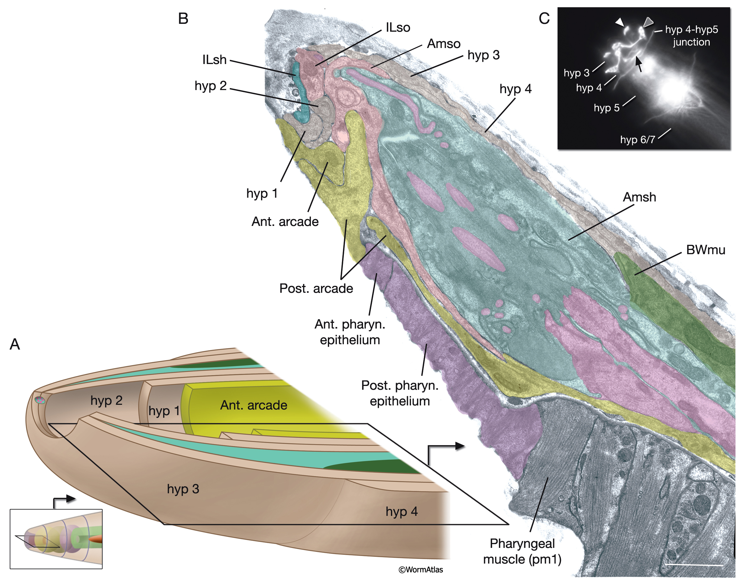
A. Three concentric rings of hypodermal cells (hyp 1, hyp 2 and hyp 3) constitute the hypodermis of the extreme anterior of the head. The innermost one, hyp 1, connects the hypodermis to the arcade cells and the pharyngeal epithelium. Not shown are posterior arcade and the pharyngeal epithelium.
B. Transmission electron micrograph (TEM) of the lateral lip, horizontal section. The endings of the head sensilla and anterior head muscles fit between the external (hyp 3 and hyp 4) and internal (hyp 1 and hyp 2) hypodermal tissues. Inside the buccal cavity, anterior and posterior arcade cells connect the buccal hypodermis to the pharyngeal epithelium. (For comparison of the positions of the anterior hypodermal and arcade cells refer to ArcFIG 2.) Color overlay has been added atop the TEM image to indicate cell types involved. Bar, 1 μm. (Image source: N533 [Hall] negative C240.)
C. Epifluorescent image of transgenic, adult-stage animal expressing the ajm-1::GFP reporter in the hypodermis, lateral view. hyp 2 and hyp 3 make adherens junctions (white arrowhead) at the tip of the lip, with the hyp 2 ring covering the interior and the hyp 3 ring covering the exterior surfaces. hyp 2 and hyp 1 rings make adherens junctions where they meet inside the buccal cavity (arrow). hyp 3 and hyp 4 rings join by adherens junctions on the outside surface (gray arrowhead). hyp 6 has already fused with hyp 7 at this stage. Original magnification, 600x. (Strain source: H. Yu and P. W. Sternberg.)
Click on picture for full resolution image.
|