|
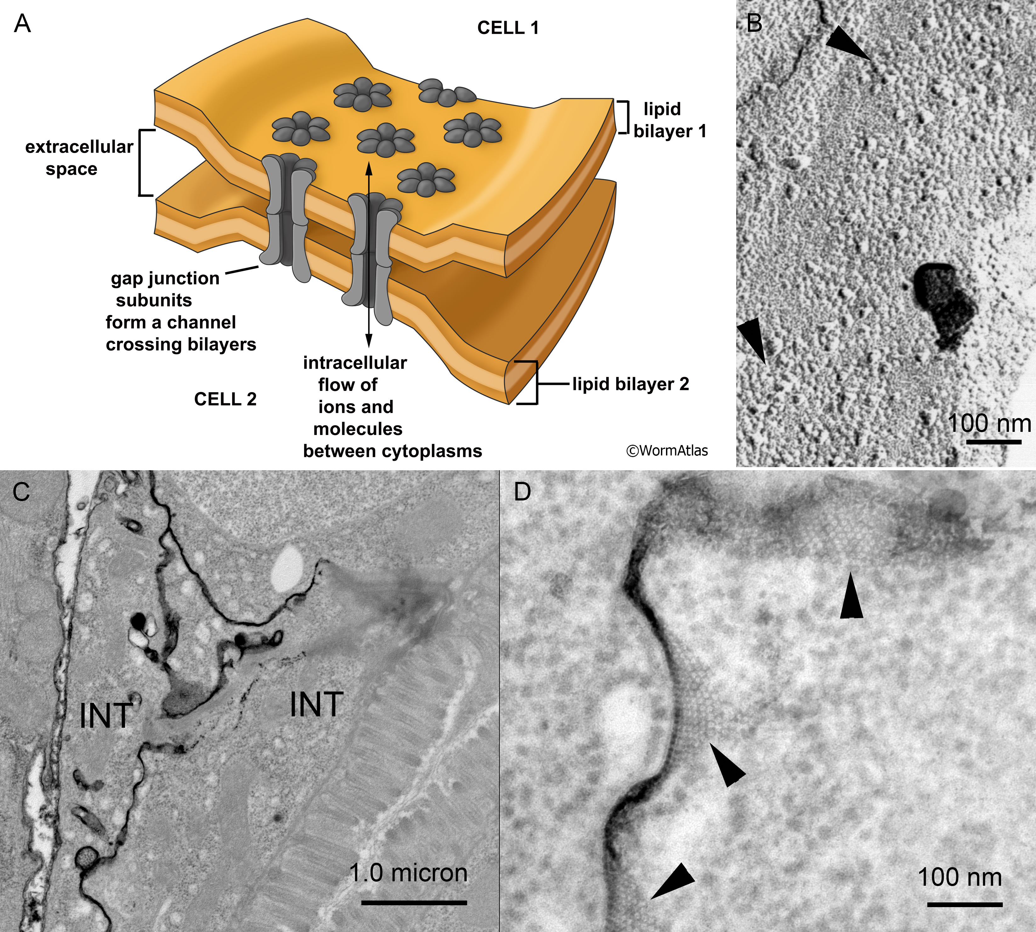
GapjunctFIG 2: Gap junction channels are clustered in the plasma membrane.
A. Schematic diagram depicting a small array of gap junction subunits lying in the plasma membranes of two closely opposing cells. The “gap” consists of the narrow space between the outer layers of the opposing plasma membranes, which is periodically spanned by the gap junction channel subunits. In a “freeze fracture replica” the fast frozen membranes are ripped open to separate the inner and outer layers of a single plasma membrane. Individual gap junction channels pull out of one layer or the other, revealing an array of intramembrane particles and pits that shows the close packing of GJ channels within the membrane, as in panel B. A typical nematode gap junction does not fracture as cleanly as for a vertebrate gap junction, where one fracture face would show all pits, and the other face all particles.
When the ionic tracer lanthanum is infiltrated into such a junction before fixation, the lanthanum will precipitate in the narrow space between the two plasma membranes. Gap junction channels will exclude this tracer as they cross the gap, leaving a negative stain that again shows the packing of the channel array as white dots against the black tracer, as in panels C & D.
B. N2 F/F Sample 4 M5 005286 (Hall) Freeze fracture replica of an adult wild type animal showing several arrays of particles and pits (arrowheads) in a hypodermal membrane where large gap junctions have formed.
C. C. elegans image 011 of 11/19/2007 (McKee). Low power view of intestine where lanthanum tracer has infiltrated between two intestinal cells (INT) at their membrane border.
D. C. elegans image 014 of 11/19/2007 (McKee). Higher power view of a nearby portion of the same sample as in panel C, with arrowheads pointing to locales where the array of GJ channels are negatively stained. Their visibility depends not only on the quality of the stain penetration, but the exact angle of the thin section vs the plasma membrane. Only where the plasma membrane is seen lying en face within the section does the particle array become visible – thus the true size of any one particle array might be much larger than what can be seen from this single section. Lanthanum tracer images are shown courtesy of Mary McKee and Emily Troemel.
Click on picture for full resolution image.
|