|
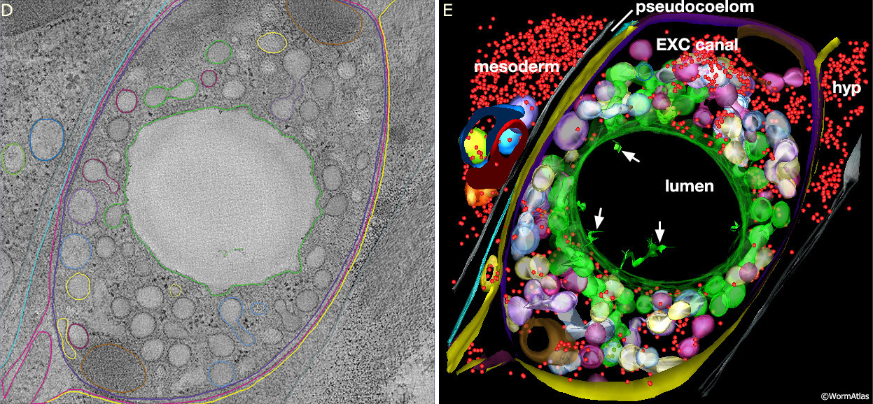
ExcFIG 7D&E: Excretory canal.
Model of the excretory canal in cross-section from an electron tomogram.
D. Orthoslice through the tomogram.
E. 3D model annotated from the tomogram.
Canal is on the right, separated from nearby mesoderm (intestine or gonad) by the narrow space of the pseudocoelom. Colors here have been chosen to mark sets of organelles by type. Red, ribosomes; green, lumen membrane and canaliculi that could be traced to luminal connections within a 400 nm interval along A/P axis (total depth of tomogram is 0.4 microns). Pale purple, white, pale yellow, blue mark other branched groups of canaliculi that must link to the lumen outside the reconstructed volume. (Purple) canal plasma membrane; (yellow) hypodermal cell plasma membrane; (brown) mitochondrion. (Arrows) indicate thin branched structures of unknown origin extending inside the excretory lumen. Note that the canaliculi are clustered close to the lumenal membrane, while ribosomes, mitochondria and other organelles are pushed to the exterior, away from the lumen of the canal. (Ashleigh Bouchelion, Kristin Politi, KD Derr, William Rice and David Hall, unpublished.)
See also ExcFIG 7A and ExcFIG 7B&C.
Click on picture for full resolution image.
|