|
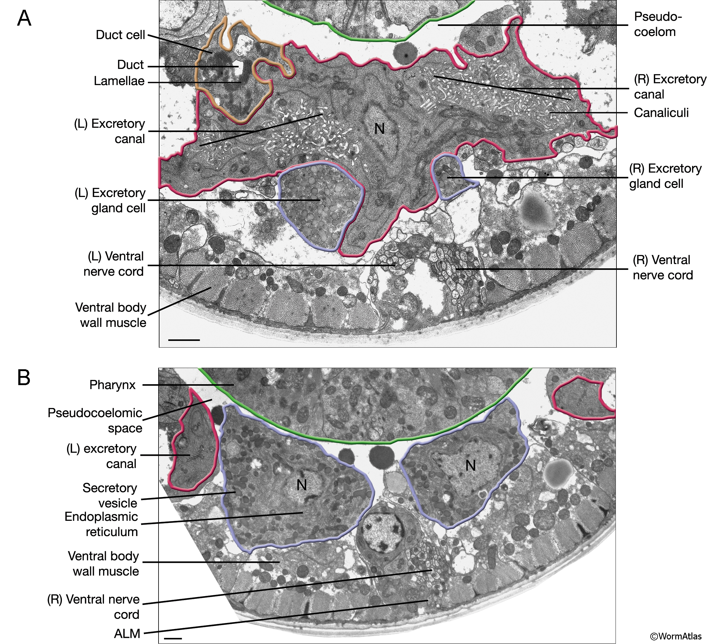
ExcFIG 4: Transmission electron micrographs (TEMs) of the excretory cell, the gland cell and the duct cell.
A. TEM of transverse section showing a high-magnification view of the excretory cell soma (red tracing) slightly posterior to the excretory sinus. The bilateral canals are still separated from each other by the large nucleus, which is located slightly to the left of the midline. The canals extending towards the midline from the lateral edges of the cell join to make one single excretory sinus in more anterior sections (not shown). The excretory cell is in contact with the pseudocoelom on the dorsal side. The left and right gland processes (blue tracings) are situated below the excretory cell and are about to be joined by a cellular bridge at the level of the secretory-excretory junction more anteriorly (not shown). The duct cell (brown tracing) is located to the left of the excretory cell and shows prominent lamella around the apical surface of the duct. (N) Nucleus. Bar 1 μm. (Image source: [Hall] N510.)
B. TEM of transverse section. The excretory gland is binucleate (blue tracing). It is located at the posterior of the head between the pseudocoelom and the ventral body wall muscle. The cytoplasm shows numerous electron-dense secretory vesicles and rough endoplasmic reticulum. (N) Nucleus. Bar 1 μm. (Image source: [Hall] N513 U4-G576.)
Click on picture for full resolution image. |