|
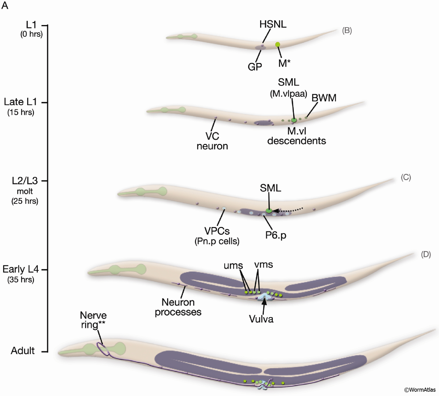
EggFIG 14A: Sex muscle development.
Schematic showing the temporal order of events leading to establishment of the adult egg-laying system, lateral view, left side. The scale on the left indicates larval stage and corresponding hours (hr) post-hatching at 20°C. Figures showing DIC/epifluorescent images of corresponding stages are indicated in parentheses on the right. For cells of the M/SM and Pnp lineages, cell nuclei only are shown. (GP) Gonadal primordium; (VPCs) vulval precursor cells. *M is located on right side of the animal. **Of the egg-laying neurons (VC1–6 and HSNL/R), only HSNL/R axons extend into the nerve ring in the adult. (Based on Sulston and Horvitz, 1977; Li and Chalfie, 1990; Garriga et al., 1993.)
See also EggFIG 14B-E.
Click on picture for full resolution image.
|