|
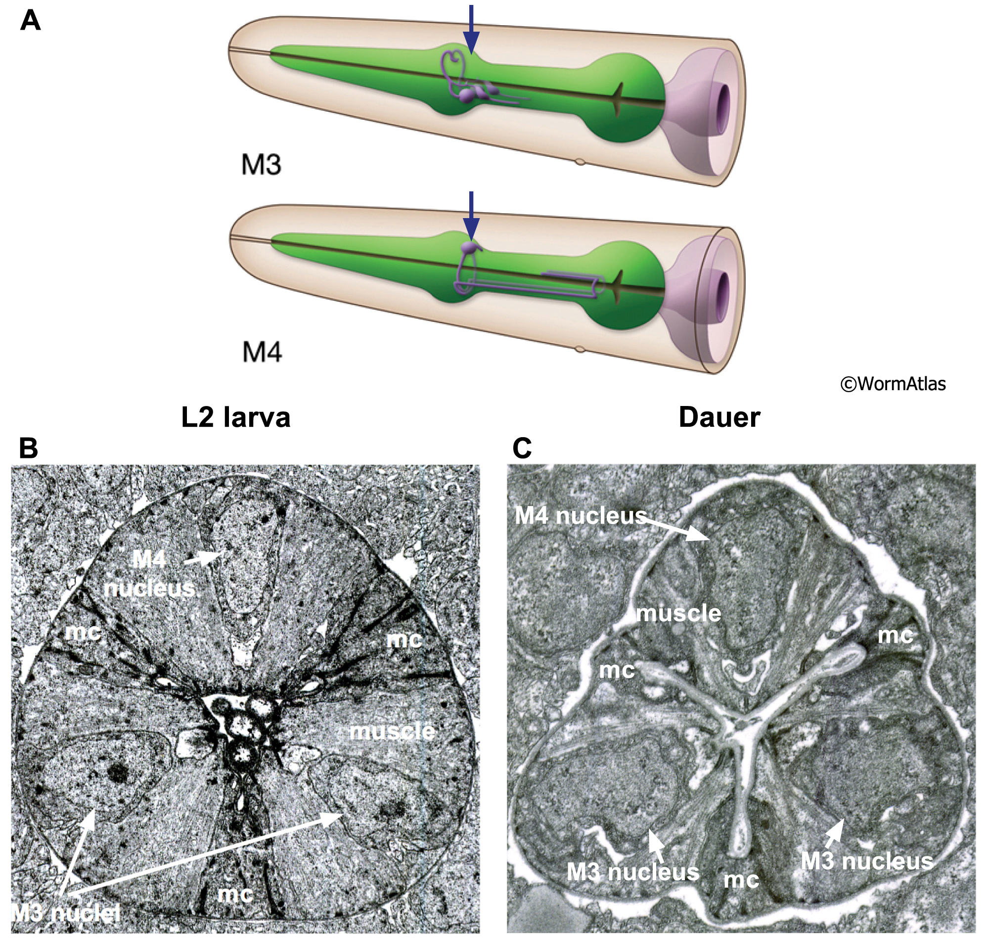
DPhaSUPFIG 8: Neuronal cell bodies in the dauer pharynx.
A. Illustrations showing the positions of the pharyngeal neurons M3 and M4. The nuclei of the M3 cell pair are located in the left and right subventral nerve cords. The M4 cell nucleus is located in the dorsal nerve cord. Arrows indicate the location of TEM sections below. (Image source: R. Ellis/WormAtlas Pharyngeal neurons.)
B. Transverse TEM of pharynx in an L2 showing the M3 and M4 nuclei and cell bodies. (Image source: N2 L2 28-14 [D. Riddle] 834.)
C. Transverse TEM of dauer pharynx showing the M3 and M4 nuclei and cell bodies. (Image source: N2 starved dauer 50-2-1 [D. Riddle] 346M.) One region of pharyngeal muscle is indicated (muscle); mc, marginal cell. The overall shape and size of the pharyngeal neuron cell bodies is preserved in the dauer, although the cytoplasmic membrane may be rougher and less smooth than in the L2. The neuron cell bodies have changed in size only modestly, if at all, while surrounding pharyngeal muscles and marginal cells have shrunken dramatically in the dauer.
Click on picture for full resolution image.
|