|
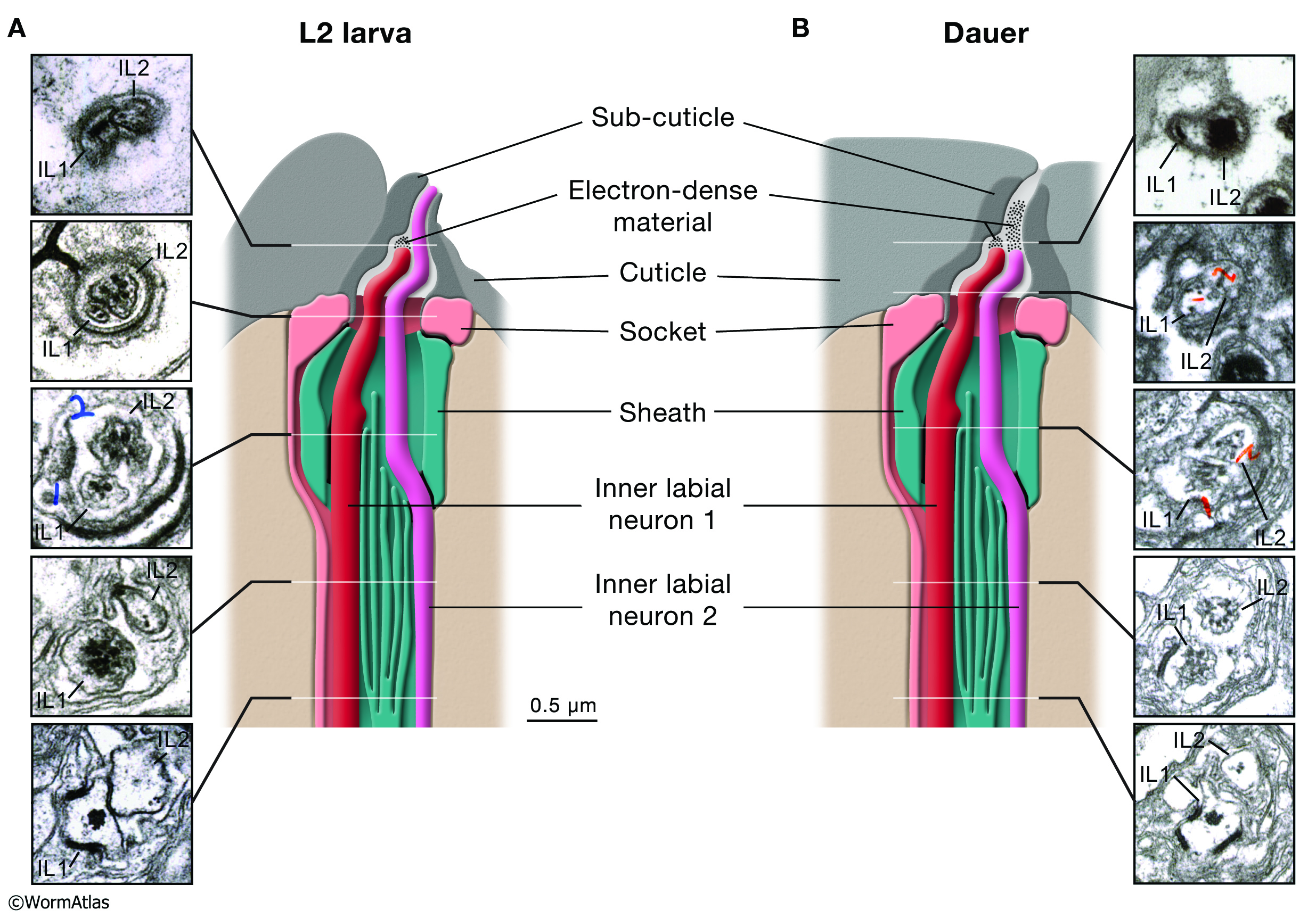
DNeuroFIG 5: Repositioning of the dauer inner labial neuron endings.
A. Left panels are transverse TEM sections of the IL channels at the approximate positions indicated on the illustration showing the positions of the IL1 and IL2 neurons in an L2 larva. (Image source: N2 L2 28-14 [D. Riddle] 6, 11, 15, 18 & 26.)
B. Left panel illustration shows alterations in IL structure in dauer larvae. Right panels show transverse TEM sections of IL channels from a dauer larva. (Image source: N2 starved dauer 50-2-1 [D. Riddle] 15, 22, 30, 39 & 44.)
The cartoons are modified from Ward et al. 1975.
Click on picture for full resolution image.
|