|
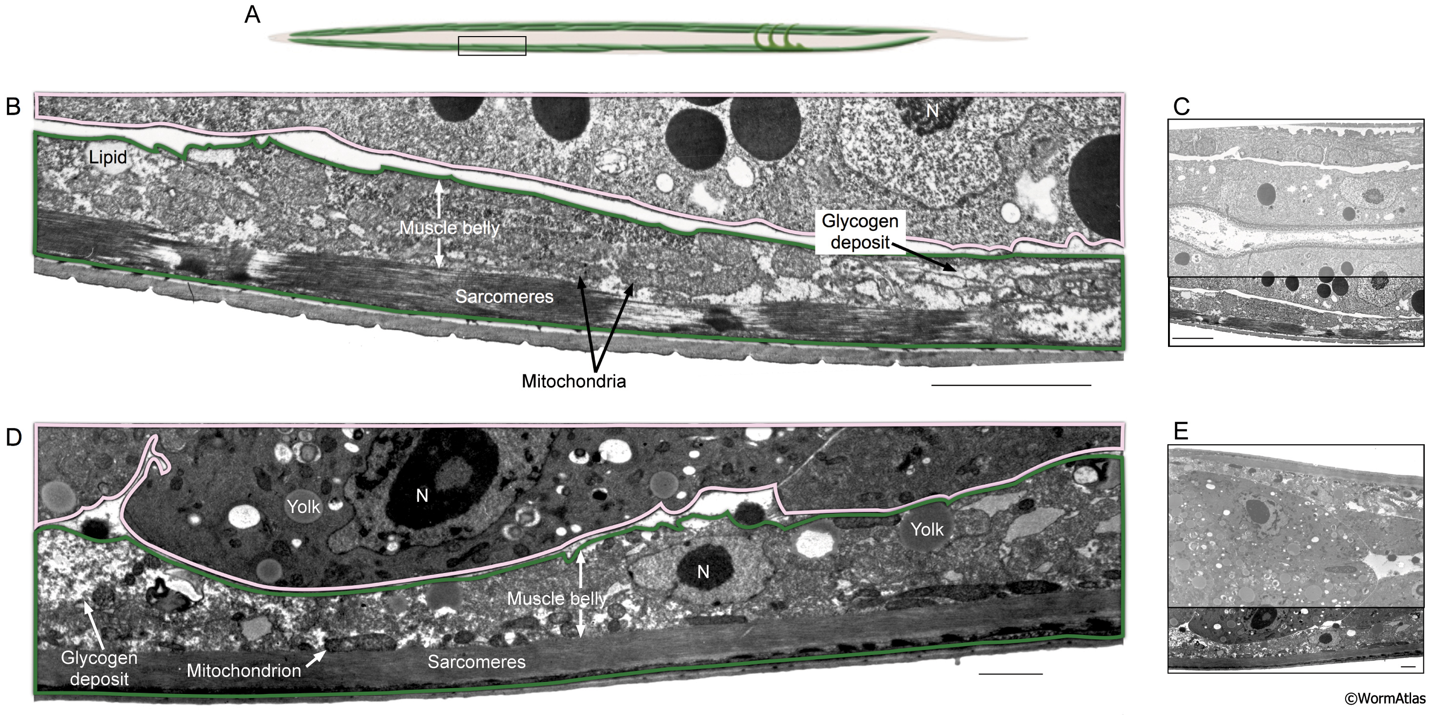
DMusFIG 4: Longitudinal view of body wall muscle in dauer and adult animals.
Longitudinal view of body wall muscles in a dauer larva and an adult.
A. Cartoon showing body wall muscle (green) with box outlining approximate position of EM sections in B-E.
B. Enlargement of dauer body wall muscle (outlined in green) showing sarcomeres, mitochondria and lipid droplets. Intestine is outlined in pink.
C. Entire TEM section shown in B, with enlarged area outlined.
D. Enlargement of adult body wall muscle (green), with intestine outlined in pink.
E. Entire TEM section shown in D, with enlarged area outlined. Lipids, yolk droplets and glycogen storage granules are shown.
N, nucleus. Bars, 5 μm. (Image sources: Dauer [D. Riddle] 56-6 109; adult [D. Hall] N533 L4 F216.)
Click on picture for full resolution image.
|