|
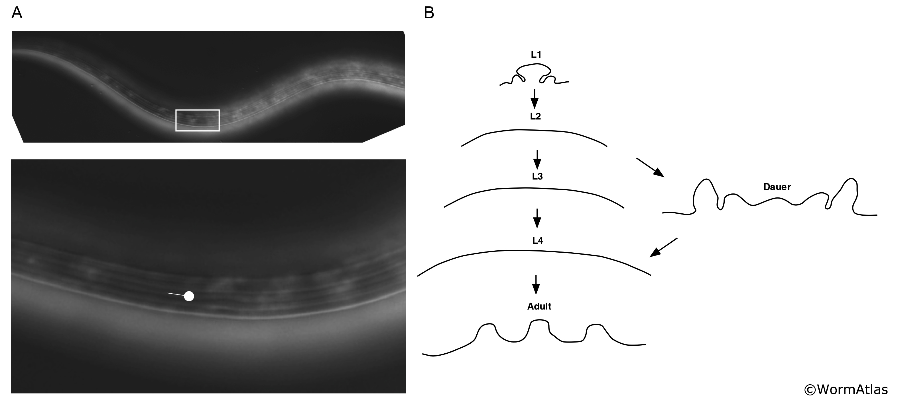
DCutFIG 2: Lateral alae in dauer larva.
A. DIC light microscopy image of dauer lateral alae. Upper panel, white box indicates magnified region below. Circle-tipped arrow lies in the central track of the alae, between the dorsal and ventral ridges. (Image source: Sylvia Lee, Cornell University.)
B. Outlines of cuticle in cross-section showing lateral alae in dauers, adults and L1s and absence of alae in L2, L3 and L4 cuticles. Dauer, L1 and adult alae profiles were adapted from Cox et al., 1981. Outlines of stages lacking alae were hand-drawn.
Click on picture for full resolution image.
|