|
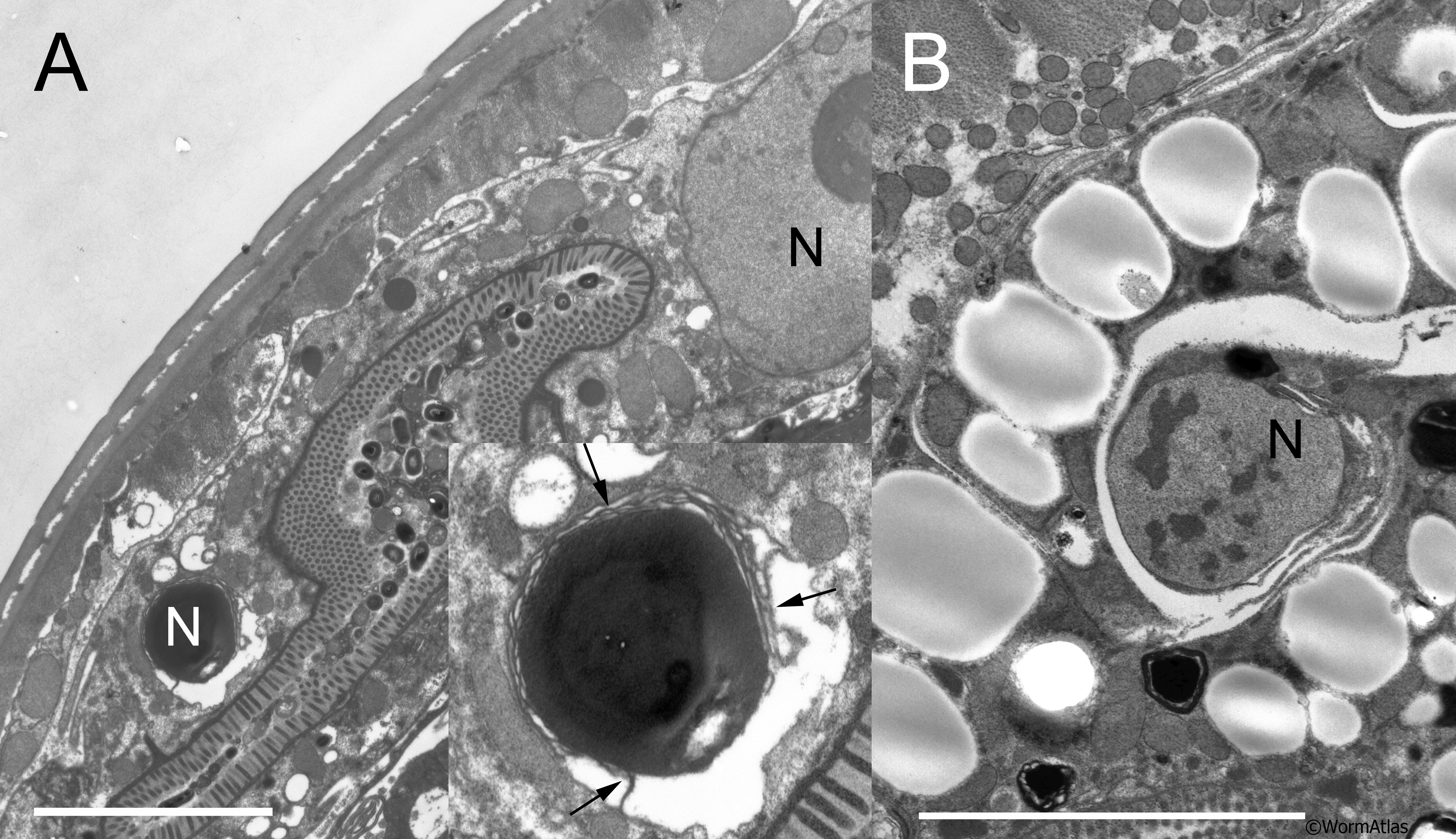
AIntFIG 9: Nuclear degeneration in aging intestinal cells.
A. A 7-day-old animal in the midbody, where the intestine has been pushed into a thin dorsal wedge by swelling of the uterus. Two intestinal nuclei are seen, a full sized nucleus on the right, with an enlarged nucleolus, and a shrunken dark-staining nucleus undergoing phagocytosis on the left, where the swollen nucleolus virtually occupies the whole volume. Inset Enlarged view of A. Arrows indicate internal membranes enveloping the shrunken nucleus. N, nucleus.
B. A nucleus from a 15-day-old animal is reduced in size and undergoing phagocytosis. Here again there is a suggestion of internal membranes beginning to envelop the shrinking nucleus, which is marked by clumping chromatin. Scale bars indicate 0.5 micron.
Click on picture for full resolution image.
|