|
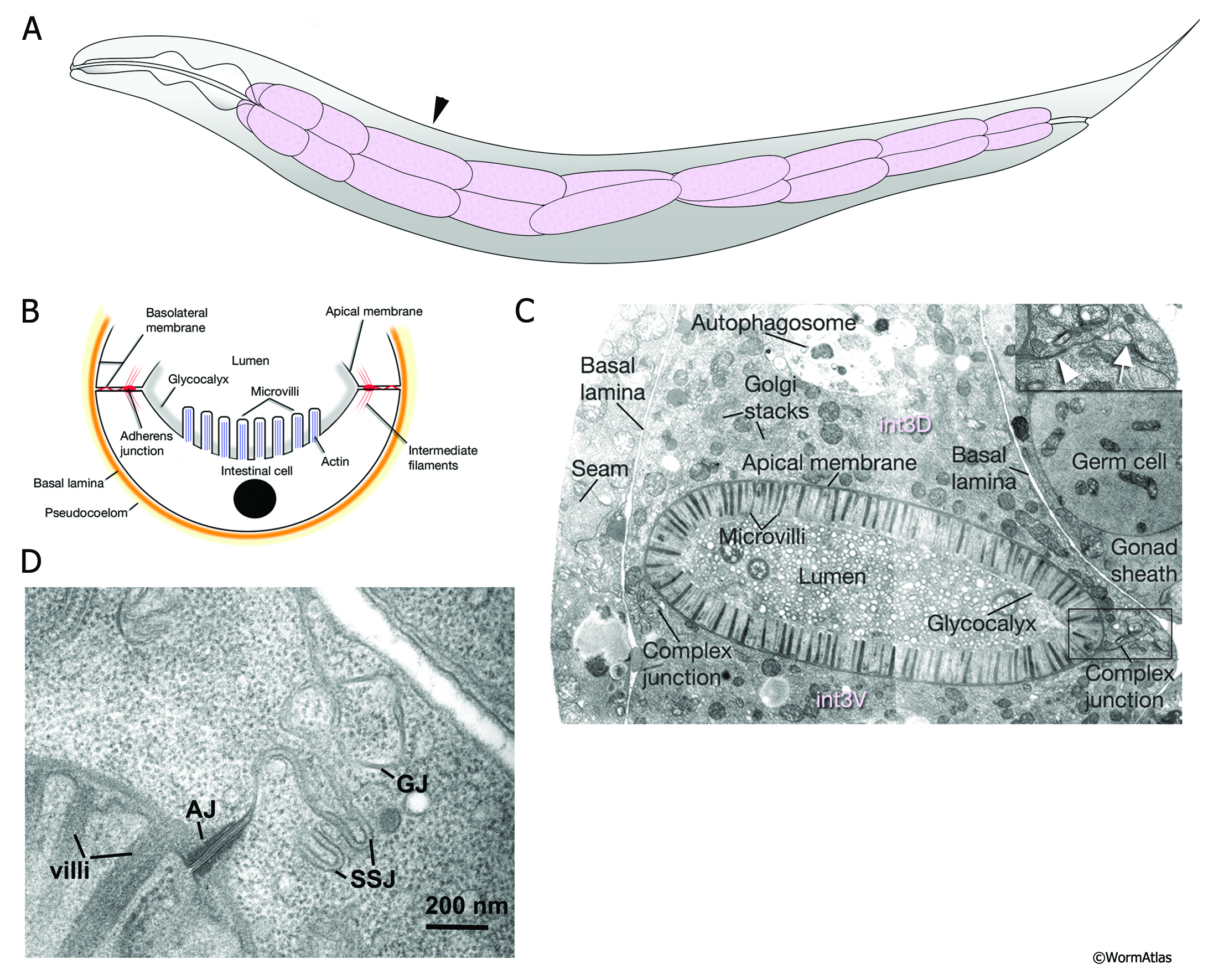
AIntFIG 1: Structure of young adult intestine.
A. The intestine is positioned on the left side of the body anterior to the vulva and on the right side of the body posterior to it. At its anterior end, the intestine is connected to the pharynx via the pharyngeal valve. The most posterior portion is squeezed by the stomatointestinal muscle (not shown), near where the intestine connects to the rectum and anus. Arrowhead indicates the position of the TEM cross-section shown in C. (Adapted with permission from Mendenhall, 2015.)
B. Key structural elements of the healthy intestinal cytoskeleton. At its basal pole the intestine is covered by a basal lamina (orange), separating it from the pseudocoelom. Pairs of intestinal cells meet to form a lumen between them, with the two cells firmly linked by adherens junctions at their apical borders. Gap junctions and smooth septate-like junctions form a complex junction just beneath the adherens junctions on the lateral membranes where the two intestinal cells meet. Intermediate filaments help to anchor a terminal web of fibers running just beneath the microvilli that face the lumen itself. An actin-based cytoskeleton fills each villus; the actin fibrils anchor into the terminal web at one end, and to an electron dense cap at the tip of the villus. A thick glycocalyx covers the outer surface of the microvilli. At adulthood, most intestinal cells contain two very large nuclei (black circle). The lumen of the young adult intestine usually is filled by debris from partially digested bacteria, but few if any intact bacteria. Graphic adapted from Wood et al. (1996).
C. Electron micrograph showing the key features of the third ring of the young adult intestine. The intestinal cytoplasm is filled with a complex mixture of organelles, including mitochondria, Golgi apparatus, RER, yolk-filled granules, and occasional large autophagosomes. A complex junction (arrow) next to an adherens junction (arrowhead) seals the two intestinal cells to each other (inset). A basal lamina covers the surface of the intestine facing the pseudocoelom. (Image source: N510 [Hall] G127.)
D. Electron micrograph showing details of the junctions connecting two intestinal cells. AJ, adherens junction; GJ, gap junction; SSJ, smooth septate junction. (Image source: N501B [Hall] 8004.)
Click on picture for full resolution image.
|