 Spermatozoon SEM
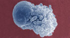
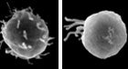
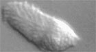
This is a mature sperm cell, or spermatozoon, from C. elegans. At the right is the pseudopod, which the cell uses for crawling. The pseudopod has knobby projections, called vilipodia, which constantly flow rearward toward the cell body.
|
 Spermatozoa Spermatozoa
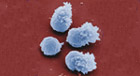
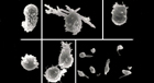
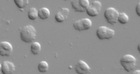
Here are four C. elegans spermatozoa. The one at left center has just started to form its pseudopod.
|
 Spermatozoon TEM Spermatozoon TEM
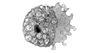
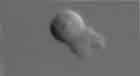
This is a cross section through a mature sperm cell, or spermatozoon, from C. elegans. At the right is the pseudopod, which the cell uses to crawl and at the left is the cell body, which contains the nucleus (dark spot at the center), mitochondria, and unusual organelles called membranous organelles.
|
 Spermatids
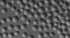
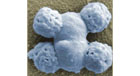
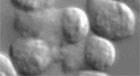
This is a picture of undifferentiated sperm cells, or spermatids, from C. elegans. Note that the cells are symmetrical, not yet having formed their pseudopod.
|
 Spermiogenesis
Scanning EM images showing the process of spermiogenesis, sperm cell differenetiation.
|
 Budding spermatids SEM
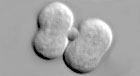
This scanning electron micrograph shows four haploid spermatids forming as products of the final cell division in spermatogenesis.
|
 Mutant spermatozoa Mutant spermatozoa
This set of SEM panels shows examples of sperm from some of the different sperm mutants. For comparison, a normal sperm is shown at the top left.
|
 Crawling sperm
Video shows C. elegans sperm crawling via its pseudopod. Note that the pseudopod, on the right, moves forward bringing the cell body of the sperm with it.
|
 Many crawling sperm
Video shows many C. elegans sperm crawling around.
|
 Treadmilling sperm
Video shows how unlike most sperm cells, which swim with a flagellum, nematode sperm crawl with a pseudopod.
|
 Spermiogenesis
Video shows spermatids undergoing spermiogenesis on a microscope slide in response to a chemical that was added to them.
|
 Dividing spermatocytes Dividing spermatocytes
Video shows two secondary spermatocytes video undergo the second meiotic division to form four haploid (i.e. having only one copy of each chromosome) spermatids.
|

|

 Click pictures for a new window with image or video and detailed legend