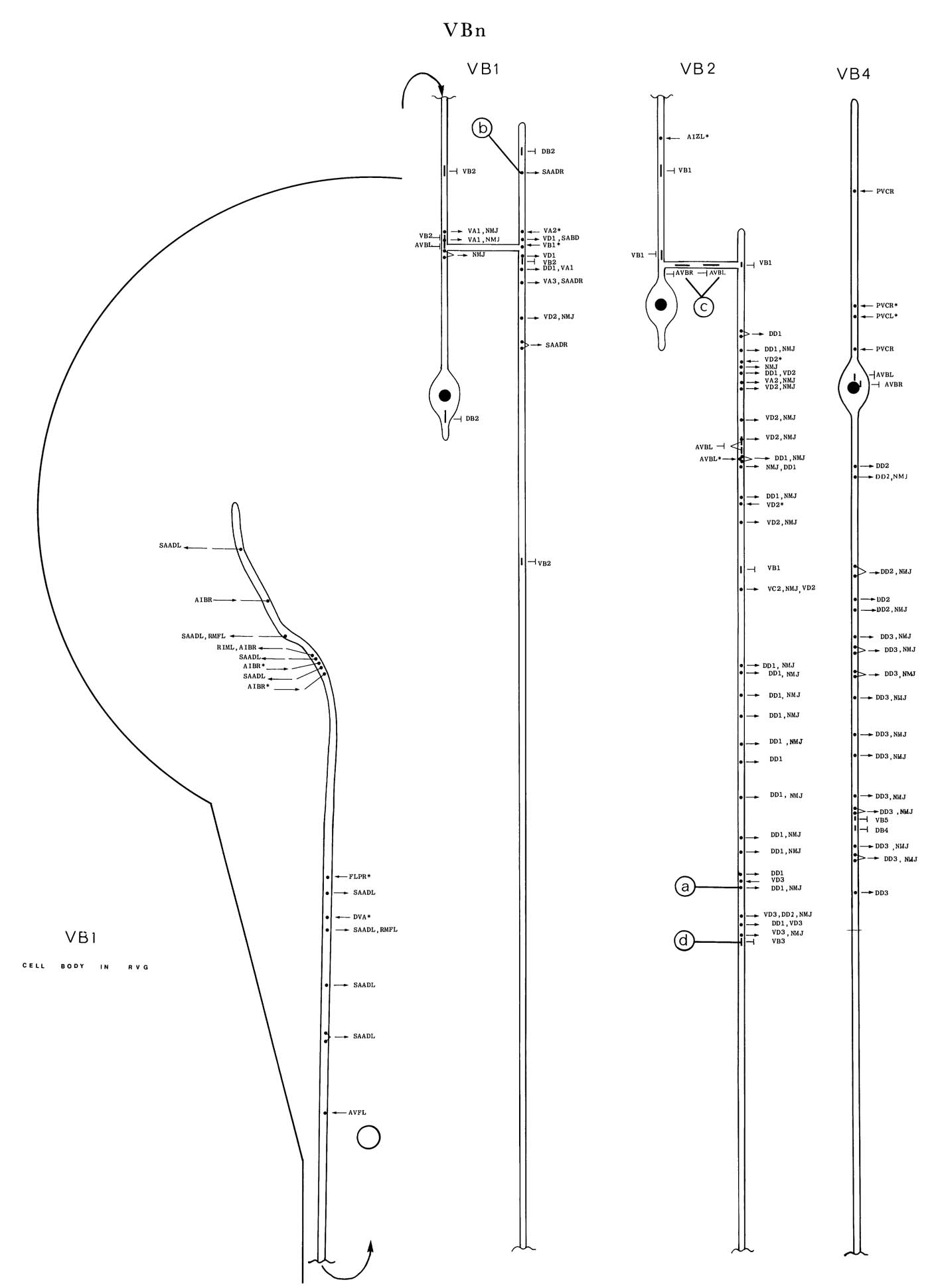
VBn is a set of eleven motoneurons, distributed along the ventral cord, which innervate
ventral body muscles. A typical VBn (e.g. VB4 (h)) has a short, anteriorly directed process
that runs in the ventral part of the cord. This process is always postsynaptic and receives
synaptic input from PVC (except in VB1 and VB2) and occasionally DVA (*d). There are
prominent gap junctions to AVB, which are usually (although not exclusively (c)) situated near
the cell body. VBn also has gap junctions with other VBns (d) and DBns;
these can be situated anywhere along the processes. A long, posteriorly directed process leaves the cell body and
moves to a position adjacent to the bounding basal lamina on the right-hand side of the cord,
next to a dorsal VAn process (figure 18). VBn processes have NMJs when they are in this
position; the NMJs are nearly always dyadic synapses with DDn neurons as the other.
postsynaptic partner (a). The posterior process of VBn eventually moves away from the NMJ
region, its place being taken by the process of the adjacent posterior VBn. The posterior distal
regions of the VBn processes then run for some considerable distance in the ventral region of
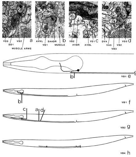
the cord, with little or no synaptic contact with other cells, before terminating. VB1 (e, f) and VB2 (g) differ in their morphology and in synaptic contacts from the other VBns. Both have
branches that run transversely across the dorsal surface of the cord just posterior of the excretory
duct, although only VB1 has NMJs in this region. VB1 also has a process that enters the left-hand side of the nerve ring, which synapses onto SAA (b) and receives some synaptic input
from AIB.
Magnifications: (a-d) x 25500.


Click pictures for higher resolution images