Members: PVR.
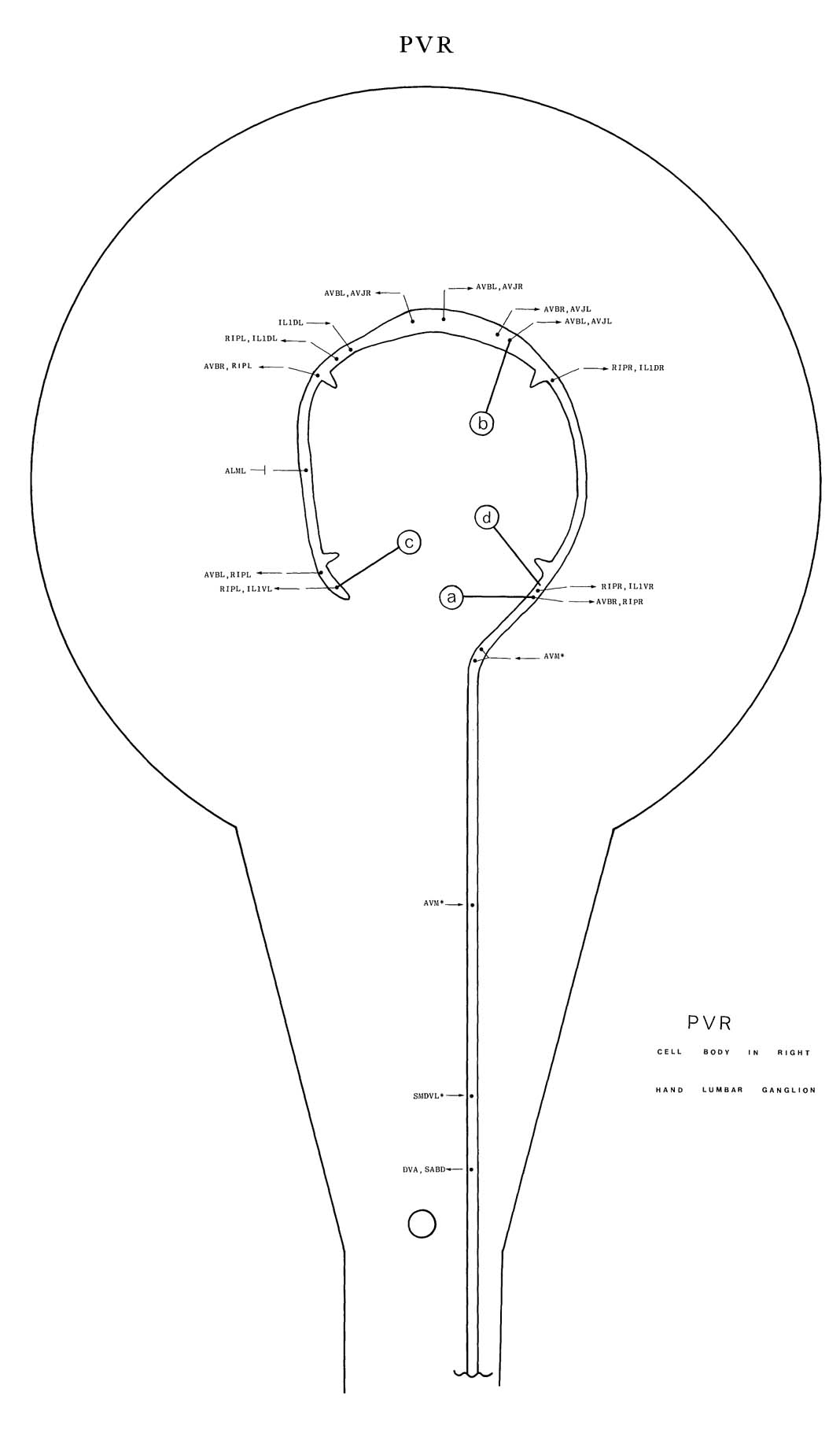
PVR has a cell body situated in the right lumbar ganglion. A posteriorly directed process
runs to nearly the end of the tailspike (e); this observation suggests that it could be a sensory
dendrite. An anteriorly directed process enters the pre-anal ganglion via the right lumbar
commissure and has a gap junction with PLMR as it crosses its process. It runs anteriorly up
the ventral cord near the ventral extremity of the process bundle. The process then enters the
right-hand side of the nerve ring and runs right round it in an anticlockwise direction, adjacent
to the inner surface of the neuropile, and ends in the left ventral region of the ring. Small
branches project anteriorly from the process at each quadrant and run in the middle of the
process bundles from the labial sensory receptors (IL1-b). No synapses are seen on these
branches. Dark-staining regions are present in the basal lamina on the inner surface of the ring,
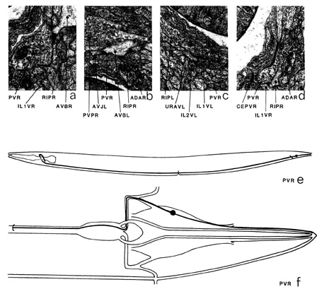
adjacent to the process of PVR, in a few places (d). The main synaptic output is in the nerve
ring and is directed to AVB (a), RIP (a, c) and AVJ (b). There is a gap junction to ALM (*d). In the ventral cord there is some synaptic input from AVM, DVA and PVM, and there
are gap junctions to DVA and PLMR.
Magnifications: (a) x 25500, (b-d) x 17000.
PVR ventral cord synapses
partners |
gap junctions |
synapses from |
synapses to and corecipients |
PDE |
- |
1m |
PDE |
PVC |
- |
1m |
1 |
AVK |
- |
- |
1 |
DB2 |
- |
- |
1 |
DB3 |
- |
- |
1 |
DA9 |
- |
- |
1 |
DVA |
2 |
1+2m |
- |
PVM |
- |
2m |
- |
LUA |
2 |
1 |
- |
PLM |
2 |
- |
- |


Click pictures for higher resolution images